Research Theme
"Developments of biological tissue evaluation using optical polarization"
 Light which propagates with rotating its polarization plane is called "Circular polarized light (CPL)".
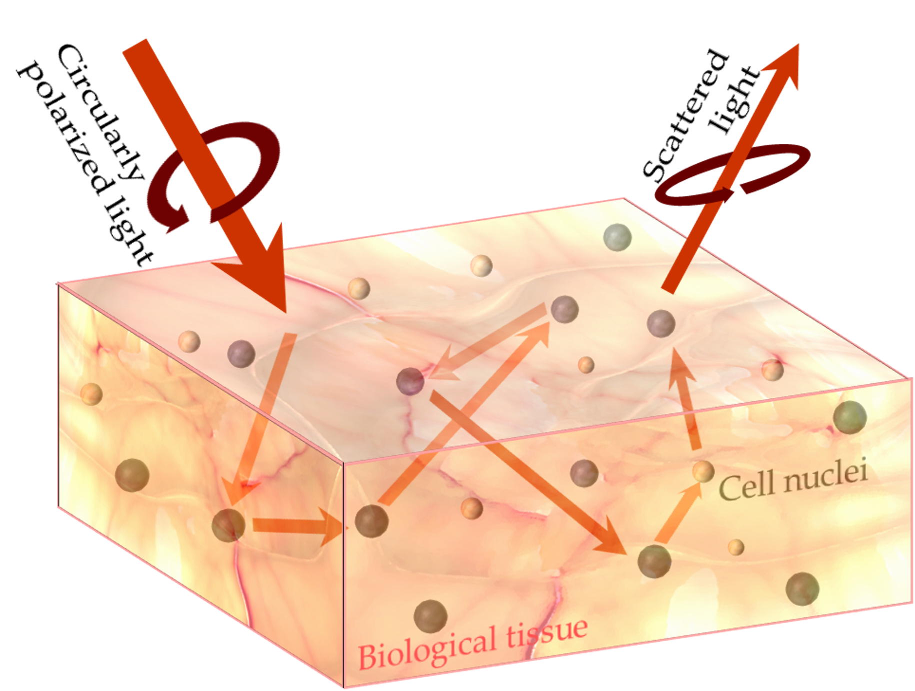
When CPL beams impinge on a biological tissue, they are scattered multiple times by cell nuclei, and depolarized gradually.
Depolarization of the resultant scattered light can provide structural information, such as the size, anisotropy, distribution, and density of the cell nucleus.
This technique, called "Circularly Polarized Light Scattering (CPLS) method" has been considered as a powerful tool for the diagnosis of tumor tissue, particularly pre-cancerous lesions. However, the loss of compact CPL sources have restricted this technique within the limited application regions.
We have achieved fully polarized CPL emission at room temperature on CPL-emitting diodes, called "spin-LEDs", which have ferromagnetic electrodes on a conventional LED structure.
The spin-LEDs integrated at the tip of an endoscope allow in vivo cancer detection using the CPL scattering technique. This technique does not require any staining, fluorescent materials, invasive ablation, and waste of time.
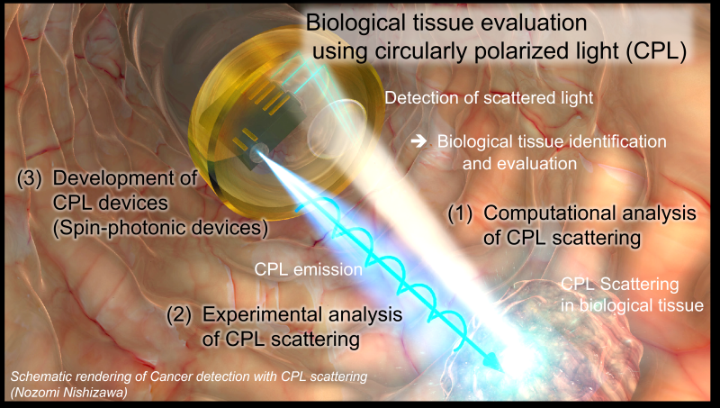
To implement this proposal, I'm studying the research from three aspects;
(1) Computational analysis of CPL scattering
(2) Experimental analysis of CPL scattering
(3) Development of CPL devices (spin-photonic devices)

(1) Computational analysis of CPL scattering
 Computational analyses using Monte Carlo method are carried out to understand deeply the scattering phenomena of CPL, which follows the analysis of (2) experimental results, the setting of the target values on (3) device development, investigations of applicable range, and (4) device designs.
The single scattering of CPL against particles with diameters corresponding to cell nuclei in healthy and cancerous tissues is investigated first. Then, we introduce them into multiple scattering systems in pseudo-healthy and cancerous tissues to clarify the contribution of optical and structural conditions to the resultant polarization of scattered light. Also, we computationally investigate the efficiency for early-staged cancer, scirrhous cancer, ulcerous colitis and the other diseases.
関連論文
- "Monte Carlo simulation of scattered circularly polarized light in biological tissues for detection technique of abnormal tissues using spin-polarized light emitting diodes"
JJAP, 59(SE), SEEG03 (2020).
(2) Experimental analysis of CPL scattering
 Experimental analysis using optical phantoms and biological tissue specimens are carried out to demonstrate the feasibility of this technique.
 Optical and structural properties of actual biological tissues are significantly complex. In contrast, optical phantom allows the investigation on an extracted parameter. Phantoms are prepared with micro-fabrication process, such as a photolithography, whose parameters can be varied systematically.
Biological tissues are prepared through collaboration research with biological and medical scientists and CPL responses are investigated experimentally.
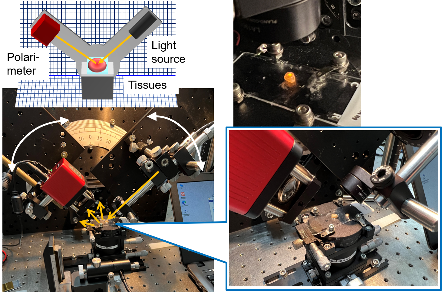
The optical setup, as shown in the figure on the right, is the double-armed rotatable systems that can change the incident and detection angles at will. A CPL source and detector is put on them, which enable to measure systematic optical geometry dependences.
Related papers
- "Angular optimization for cancer identification with circularly polarized light"
Journal of Biophotonics, 14, (3), 202000380 (2020).
Press Rerease 東工大ニュース Tokyo Tech News
(3) Development of CPL devices (spin-photonic devices)
 Spin-polarized electrons injected from ferromagnetic electrodes into semiconductor LED structure are combined with holes, resultantly generate light having spin polarization, CPL. This device is called as a "spin-LED". The TITECH group to which I belonged have studied Spin-photonic devices represented by spin-LED. We achieved fully polarized CPL emission at room temperature, electrical switching of CPL helicity at high speed, and detection of CPL at room temperature.
CPL cannot be transferred through a tortuous optical fiber, while maintaining its polarization. However, the spin-LED can directly emit, control, and detect CPL even at spatially restricted places. Combined with the above-mentioned CPLS technique, novel in vivo biological tissue evaluations can be developed.
I progress the development the spin-photonic devices suitable for biological applications; suitable wavelength, higher speed switching of CPL helicity, and so on.
Spin-polarized electrons injected from ferromagnetic electrodes into semiconductor LED structure are combined with holes, resultantly generate light having spin polarization, CPL. This device is called as a "spin-LED". The TITECH group to which I belonged have studied Spin-photonic devices represented by spin-LED. We achieved fully polarized CPL emission at room temperature, electrical switching of CPL helicity at high speed, and detection of CPL at room temperature.
CPL cannot be transferred through a tortuous optical fiber, while maintaining its polarization. However, the spin-LED can directly emit, control, and detect CPL even at spatially restricted places. Combined with the above-mentioned CPLS technique, novel in vivo biological tissue evaluations can be developed.
I progress the development the spin-photonic devices suitable for biological applications; suitable wavelength, higher speed switching of CPL helicity, and so on.Related papers
- Reveiw Paper "Lateral-Type Spin-Photonics Devices: Development and Applications"
Micromachines, 12(6), 644 (2021).
- "Pure circular polarization electroluminescence at room temperature with spin-polarized light-emitting diodes"
PNAS, 114(8), 1783-1788 (2017).
Press Rerease 東工大ニュース Tokyo Tech NewsSeiichi Tejima Research Award (Paper Award)
- "CIRCULARLY POLARIZED LIGHT-EMITTING DIODE" (Japan patent JP2020-136576)
- "DOMAIN WALL MOVEMENT TYPE SPIN EMITTING DEVICE" (Japan patent JP2015-41690)
- "DUAL ELECTRODES TYPE SPIN EMITTING DIODE AND LASER" (Japan patent JP2014-120692)
(4) Device develop for biological tissues evaluations
 Integrating these studies, we develop the CPL subassembly for biological tissue evaluations.
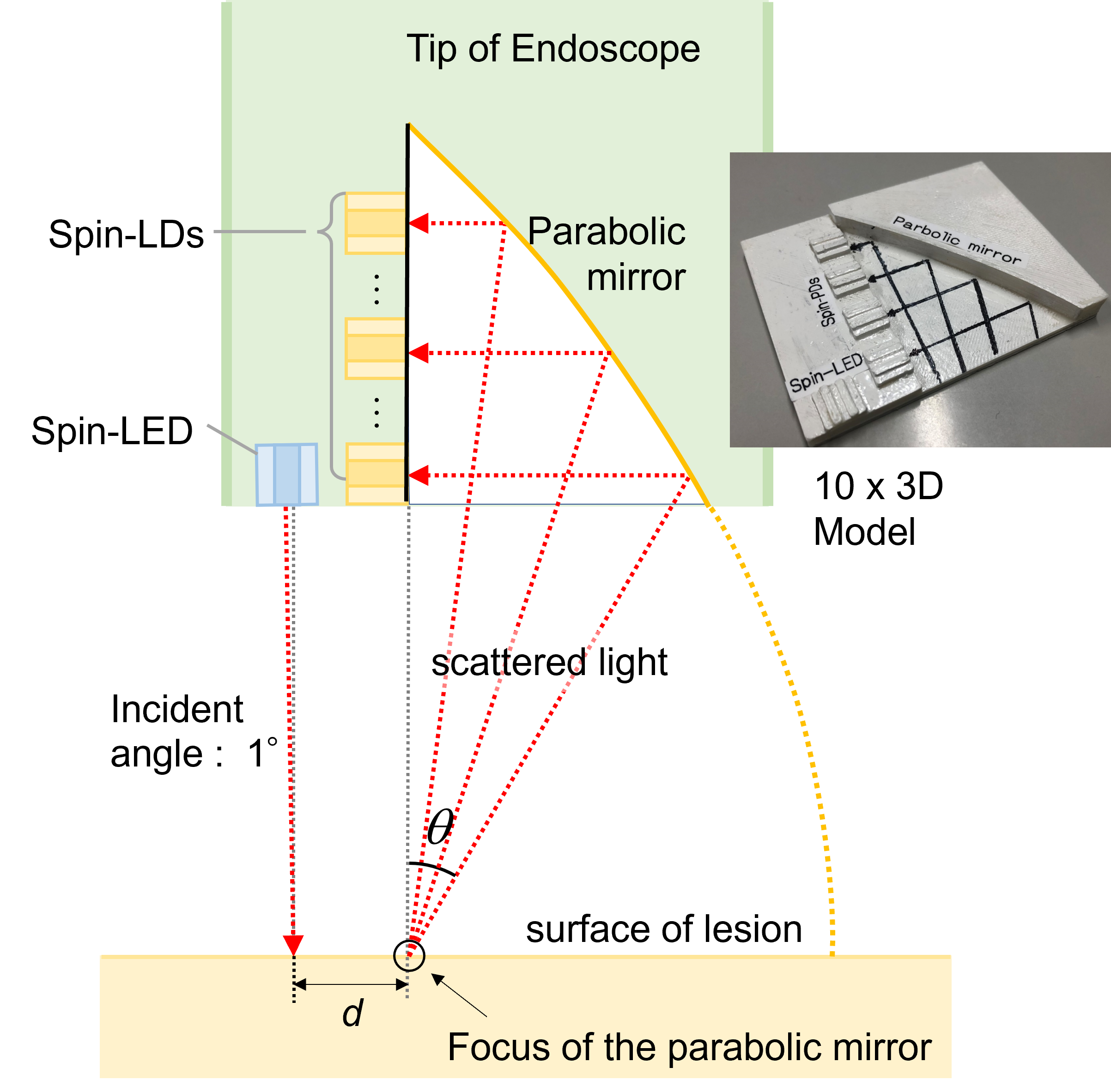
From our previous study, the sampling depth of the biological tissues can be modulated by tuning the detection angle. Therefore, the depth profile in tissues can be obtained from the detection angle dependence of scattered light.
I designed the structure of the endoscope probe consisting of spin-photonic devices and a parabolic mirror, which can detect the scattered light with different detection angles simultaneously(Patent pending).
Presently, I'm studying a one-tenth model of this subassembly, as shown in figure.
 Patents
- "ENDOSCOPE PROBING DEVICE" (Japan patent JP7352251)


